
Management of Infantile Spasms in Infants Under One Year of Age
| Written Oct 2016 | Dr Tekki Rao, Dr Alasdair Parker & Dr Deepa Krishnakumar |
| Updated Sep 2021 | Dr Mayy El Gamal, Dr Manal Issa & Dr Pooja Harijan |
| Updated Jul 2025 | Dr Tekki Rao, Dr Sadia Bashir & Dr Sandra Walton-Betancourth |
| Next review | July 2028 |
This guideline is for the use of general paediatricians, trainees, and paediatric neurologists. Any specific cases in which there is diagnostic uncertainty should be discussed with the tertiary paediatric neurology team.
Click each section to expand/collapse it.
Download a PDF copy of this guideline
Background
Background
Infantile spasms (IS) are the most common severe infantile epilepsy, characterized by clinical spasms that significantly impact the development of affected children. Recently, the term “Infantile epileptic Spasm Syndrome” (IESS) has been proposed by ILAE (International League Against Epilepsy) to encompass both West syndrome as well as infants presenting with epileptic spasms (ES) who do not fulfil all criteria for West syndrome.
West syndrome:
WS is regarded as a subtype of ISs and is the most frequently reported subtype presenting in about 90% of cases of Infantile spasms. The disorder mainly affects those in their first year of life.
West syndrome” is classically defined by the triad of ES, hypsarrhythmia, and developmental stagnation or regression. The spasms present with characteristic EEG changes known as hypsarrhythmia and a strong association with developmental delay or regression.
Epileptic spasms:
ES are mandatory for diagnosis and consist of a sudden flexion, extension, or mixed extension–flexion of predominantly proximal and truncal muscles that is usually more sustained than a myoclonic jerk. ES usually occur in series or clusters, often on awakening, with an increasing prominence of motor features through the cluster, often over minutes, though clusters may last 30 minutes or longer. ES may be symmetric or asymmetric. In addition, symptoms can occasionally be subtle (e.g., serial tonic contraction of single muscles or muscle groups, grimacing, head nodding, chin movements, eye movements) and may only be identifiable on ictal electroencephalography (EEG) recordings. Non-serial, single ES may also be observed but should only be attributed to ISS (“infantile spasm single variant”) when the ictal or interictal EEG is compatible. It should be noted that focal seizures may also co-occur independently of spasms or even preceding spasms, especially in a structural aetiology, such as tuberous sclerosis complex (TSC).
Classification:
Infantile spasms can be classified into 3 groups by ILAE:
- Idiopathic/(Presumed) Genetic
- Symptomatic/structural /metabolic/identified aetiology:
- Cryptogenic/Unknown aetiology.
Aetiology
Aetiology
Every effort should be made to determine the aetiology of the IS as this can help determine the likelihood of response to treatment, guide therapeutic decisions and help provide a more definitive prognosis for the child. However, this should not delay the start of treatment once the diagnosis is confirmed.
A. Symptomatic/structural/metabolic/identified aetiology:
The identified etiology is found in 60% to 70% of symptomatic infantile spasms cases, and can be divided into prenatal, perinatal, and postnatal.
Prenatal
- CNS malformations: Cortical dysplasia, Cerebral dysgenesis, lissencephaly, holoprosencephaly, and hemimegalencephaly account for the additional CNS malformations associated with infantile spasms.
- Neurocutaneous disorders: The most common neurocutaneous disorder associated with infantile spasms and accounting for 10% to 30% of prenatal causes is tuberous sclerosis complex (TSC), in which 68% of patients have infantile spasms. The other neurocutaneous disorders with etiological associations to infantile spasms are nevus linearis sebaceous, incontinentia pigmenti, Ito syndrome, and neurofibromatosis type 1.
- Chromosome abnormalities: Down syndrome is the most common chromosomal abnormality associated with infantile spasms. Up to 15% of prenatal causes of infantile spasms are attributed to chromosome abnormalities, including 18q duplication, 7q duplication, deletion of the MAGI2 gene on chromosome 7q11.23-q21.11, and partial 2p trisomy.
- Genetic mutations: In addition to chromosomal abnormalities, genetic mutations such as those encoding the forkhead protein G1, syntaxin-binding protein 1, calcium or calmodulin-dependent serine protein kinase, ALG13, pyridoxamine-5’-phosphate oxidase, and adenylosuccinate lyase are associated.
- Inborn errors of metabolism: Twenty-five metabolic disorders have associations with infantile spasms. Phenylketonuria is the most common inborn error of metabolism with etiological associations to infantile spasms in countries where PKU is not identified at birth; this accounts for 12% of patients with PKU.
- Congenital infections: Congenital infections are the last prenatal insult that must be considered with associations to infantile spasms. These congenital infections include toxoplasmosis, syphilis, cytomegalovirus infection, and Zika virus infection.
Perinatal:
Though prenatal factors account for the greatest proportion of cases of symptomatic infantile spasms, perinatal causes of infantile spasms, including hypoxic-ischemic encephalopathy and neonatal hypoglycaemia.
Postnatal:
These include traumatic injury, near drowning, tumors, and CNS infections with attribution of 15% to 67% of cases of symptomatic infantile spasms.
B. Cryptogenic/ No identified aetiology.
Cryptogenic infantile spasms have no identifiable cause and the following criteria: no other kind of seizures, a normal examination, a normal CT and MRI, recurrence of hypsarrhythmia between consecutive spasms of a cluster, and lack of any focal interictal or ictal EEG abnormalities. 10% to 40% of patients are classified as cryptogenic. Cryptogenic infantile spasms are associated with a better prognosis as compared to symptomatic infantile spasms.
C. Idiopathic/(Presumed) Genetic
ILAE has proposed an additional group to differentiate a subset of cryptogenic infantile spasms based on the presence or absence of developmental delay before the onset of symptoms, identified as idiopathic.
Establishing the Diagnosis
Establishing the Diagnosis
- Obtain a thorough history:
- History consistent with Infantile spasms (symmetric or asymmetric)
- Directly witnessed event /video of seizures seen by a senior clinician
- Antenatal and neonatal history, including history of assisted conception and country of birth ( due to variations in newborn screening programs and infectious diseases ).
- A thorough family tree should be constructed, with special attention to consanguinity, ethnicity, and family history of neurological disorder and/or genetic disorder.
- Careful attention should be given to the child’s past and present developmental status (onset of spasms is often associated with plateauing or regression of development)
- Clinical assessment:
- Look for dysmorphic features, growth parameters, head circumference should be plotted, and evidence of IU infections (microcephaly, hydrocephalus, retinitis, petechiae and hepatosplenomegaly/jaundice)
- Complete neurological examination and CVS examination for comorbidities like (Down syndrome, or TS)
- Look for neurocutaneous stigmata and actively look for tuberous sclerosis / NF1 (including wood’s lamp examination)
- Assess development – document pre-morbid developmental delay
- Refer for ophthalmology for fundus examination
- Abnormal clinical signs should initiate specific referrals such as:
- Cutaneous stigmata or dysmorphism – referral to a geneticist.
- Spleno/hepatomegaly – referral to a metabolic specialist
- Referral to clinical genetics is recommended in all cases where an acquired cause is unlikely.
- EEG:
- The EEG should get a full sleep-wake cycle and a full ictal event, best obtained with an overnight inpatient 24-hour video EEG. If the diagnosis is unclear on the initial EEG, repeat or prolonged monitoring can be performed 1 to 2 weeks after the initial study.
- Classical/modified hypsarrhythmia or compatible EEG findings (No consensus exists on the exact EEG abnormality required to establish the diagnosis)
- If clinical or EEG findings are uncertain – discuss with Paediatric neurologist, Addenbrookes Hospital
Further investigations
Further investigations
Table 1: 1st and 2nd line investigations
| First Line | Second Line |
|---|---|
| Electrophysiology Urgent EEG Sleep / Ictal EEG (if not achieved already) | Electrophysiology Review/Repeat Sleep / Ictal EEG For prolonged EEG if still diagnostic uncertainty |
| Biochemical Full blood count, U+E, LFT, blood gas/bicarbonate, glucose, calcium, magnesium, ammonia, plasma lactate | Biochemical Serum TFT, Uric acid, Plasma transferrin glycoforms, Very long chain fatty acids, Urine sulfocysteine, Plasma amino acids, Biotinidase, Acylcarnitines, Urine organic acids, Urine and plasma creatine and guanidinoacetate CSF: glucose, lactate, amino acids (with paired plasma samples) |
| Imaging MRI brain 1.5T NB: recommended in all children, even where the aetiology is clear on clinical grounds | Imaging Repeat 1.5T if original poor quality or child < 3 years MRI brain 3T |
| Genetic Array CGH Urgent trio WGS (R14) Clinical genetics opinion For further information, click here | Genetic Discuss with clinical geneticist, when Trio WGS negative and genetic cause is suspected |
Before Treatment
Before Treatment
- Provide information about infantile spasms to the parents
- Discuss treatment options including side-effects with parents
- (Prednisolone, ACTH, Vigabatrin alone or in combination). Discussion should be carried out with parents about all pros and cons of combination therapy, including lack of evidence for improved developmental outcome or relapse rate with its use.
- For steroid treatment:
- Document baseline blood pressure
- Are there any contraindications for steroid?
- Rule out active bacterial infection (clinical +/-investigations)
- Give the parents and carers a steroid card and information about when to seek medical help for side effects.
Aim of successful treatment
Aim of successful treatment
Early diagnosis and shorter lead time to treatment is the gold standard to get an effective response which is measured by: freedom from IS, usually after a treatment interval of not more than 2 weeks, and without relapse of seizures over a pre-specified interval such as 1–3 months.
The aim of the treatment should be the rapid cessation of epileptic spasms and the rapid resolution of hypsarrhythmia on the EEG (both awake and asleep) as a prerequisite for the best possible cognitive development of the affected child.
Treatment
Treatment
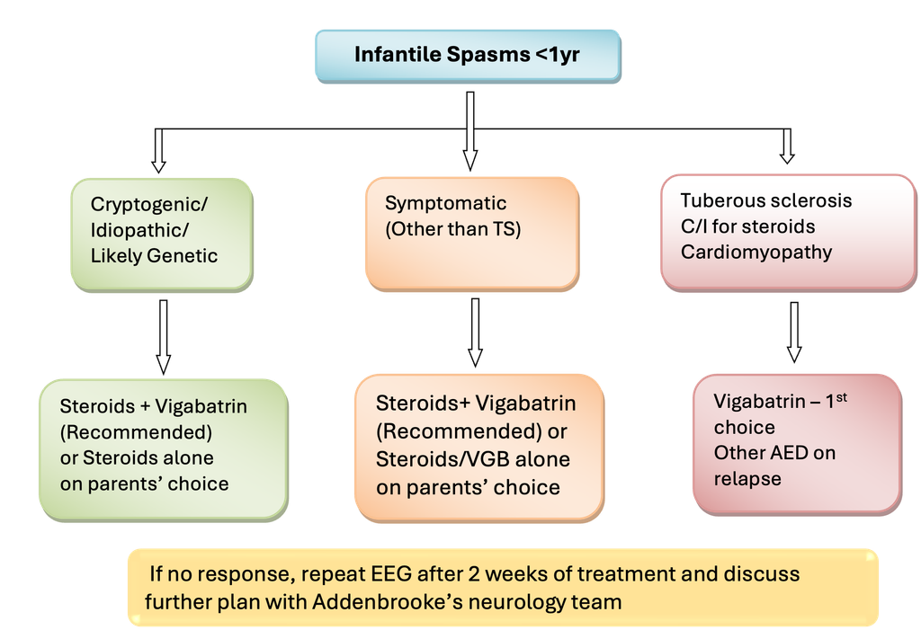
NICE recommendation (Jan 2025):
First-line treatment:
- Offer combination therapy with high-dose oral prednisolone and vigabatrin as first-line treatment for infantile spasms that are not due to tuberous sclerosis, unless the child is at high risk of steroid-related side effects.
- Consider vigabatrin alone as first-line treatment for infantile spasms in children at high risk of steroid-related side effects.
- Offer vigabatrin alone as first-line treatment for infantile spasms due to tuberous sclerosis. If vigabatrin is ineffective after 1 week, add high-dose oral prednisolone.
Please contact Paediatric Neurology Registrar at Addenbrookes on 01223 245151 pager 157488 or CDC on 01223 216662.
STEROID TREATMENT
STEROID TREATMENT
(Excluding children < 5kg or younger than 2 months)
Treatment from Day 1 to Day 14 (Prednisolone or ACTH – not both)
Oral Prednisolone:
- 10mg per dose, four times a day for 14 days.
- If spasms continue on Day 7 or reappear between Day 8 and Day 14 inclusive, increase the dose to 20mg per dose three times a day for the remaining doses.
Weaning Steroid Treatment after Day 14 in those with no spasms
If receiving Prednisolone 10mg four times a day, titrate the dose as:
- 30 mg daily for 5 days
- 20mg daily for 5 days
- 10mg daily for 5 days
- 5mg daily for 5 days
- 2.5mg daily for 5 days
- 1 mg daily for 5 days then Stop
If receiving Prednisolone 20mg three times a day on Day 14 titrate the dose as :
- 40 mg daily for 5 days then
- 20mg daily for 5 days and finally
- 10mg daily for 5 days then stop
- 5mg daily for 5 days
- 2.5mg daily for 5 days
- 1 mg daily for 5 days then Stop
OR
Tetracosactide (ACTH):
- Intramuscular injection using depot injection, 500 micrograms once daily on alternate days for 3 days in a week (Mon, Wed, Fri).
- If spasms continue on Day 7 or reappear between Day 8 and Day 14 inclusive, increase the dose to 750 micrograms once daily on alternate days for the remaining doses.
ACTH injection should be given by health care professional who can deal with allergic reactions and in a hospital setting. Keep necessary equipment and drugs to deal with anaphylaxis. Watch for reactions such as marked redness, pain at reaction site, urticaria, pruritus, flushing and dyspnoea. Switch to prednisolone, if reactions are noted.
Weaning of ACTH at Day 14:
If receiving 500 micrograms during first 2 weeks:
- 250 micrograms IM on alternate days (Mon, Wed, Fri) – week 3
- 250 micrograms IM twice in week (Mon, Thu) – week 4
- 250 micrograms IM once in a week – week 5
- then stop
If receiving 750 micrograms during first 2 weeks
- 500 micrograms IM on alternate days (Mon, Wed, Fri) – week 3
- 250 micrograms IM on alternate days (Mon, Wed, Fri) – week 4
- 250 micrograms IM twice in week (Mon, Thu) – week 5
- 250 micrograms IM once in a week – week 6
- then stop
Monitoring steroid treatment
- Please add omeprazole/lansoprazole until end of treatment (to avoid gastric irritation).
- Monitor blood pressure every 3 days (At least twice in a week) for 14 days. Monitoring can be discontinued during weaning unless elevated at the end of 14 days. If elevated on day 14 monitoring should be continued until stopping treatment and blood pressure returns to normal range after stopping treatment.
- Treat elevation in blood pressure above the 95th centile for age (usually above 120/90) with thiazide diuretic or nifedipine as per BNFC and monitor 48 hourly thereafter until acceptable, being prepared to increase antihypertensive dose as necessary.
- Provide urine dipsticks and ask to check urine sugar at 48 hours and then weekly till end of treatment.
- After 3 days treatment with prednisolone or 2 doses of ACTH, consider child to be immunosuppressed and treat fevers as per local febrile neutropenia protocol.
- Counsel regarding chicken pox exposure – consider zoster immune globulin if exposed and treat with IV Aciclovir if vesicles appear whilst on treatment. The preventive effect of VZIG lasts for about three weeks. Antibodies degrade after that time. If there is a new contact (after three weeks) or continued contact from the same index patient (household?), VZIG will be needed again after three weeks. The dose for oral Aciclovir prophylaxis is 40mg/kg/ day in four divided doses for one week, starting from day seven of contact (contact only once) If there is continued contact start Aciclovir from day 7 till the end of contact + 7 more days.
- Arrange open access to paediatric ward for acute febrile illnesses during first 6 weeks.
- Adrenal Suppression Monitoring: Check for early morning cortisol level, 1 day after stopping treatment with steroids. Take action as per the level
- Level >300 nmol/L – No action
- Level 150-300 nmol/L – Repeat test another day, if still low refer to local endocrinology colleagues
- Level <150nmol/L – supplement with oral hydrocortisone (Weight <5kg : 0.5mg QDS, weight 6-10kg :1mg QDS, weight 11-13kg: 2mg TDS, weight 14-20kg: 2.5mg TDS). Also please consult local endocrinologist.
- Consider the child to have adrenal suppression for 6 weeks after the end of treatment and consider need for hydrocortisone replacement if the child develops an acute illness during this period.
VIGABATRIN TREATMENT
VIGABATRIN TREATMENT
- Route: Oral
- Dose:
- Day 1 – 25mg/kg/dose, twice a day
- Day 2 – 50mg/kg/dose, twice a day
- If no spasms: continue the same dose until weaned.
- If spasms continued until day 4 or reappeared between Day 5 and 14, increase the dose to 75mg/kg/dose twice a day before day 14. (Max daily dose of 3 g/day to avoid visual field defects)
- Practical information: Each sachet of 500 mg of Vigabatrin is made up in 10 ml of water making a mixture containing 50 mg per ml water. The dosage will be given to the nearest 25 mg dose (0.5 ml), i.e. round up or down to the nearest 0.5 ml. NG administration: Sachet contents dissolve completely in 10 mL of water and flush down an 8Fr NG tube without blockage.
- Duration of treatment: 3 months, withdraw at 3 months (Dose adjusted as per weight gain, increasing as the body weight increases, in increments of 25 mg per dose (50 mg per day) as required until 3 calendar months from Day 0). Duration may be less than 3 months on clinician’s discretion when early remission is observed.
- Monitoring: Watch for drowsiness and confusion, excitation and agitation can also happen. Feeding failure may occur as well, and may require NG tube feeds. Visual field defects are not possible to monitor. Renal failure: dose should be reduced if creatinine clearance is less than 60 ml/minute/1.73 m2.
- Weaning: Gradual reduction in 4 weeks and then stop.
Combination treatment
Combination treatment:
Doses, escalation and weaning with both agents would be same as for individual agents, which, means 2 weeks of steroids and 3 months of Vigabatrin followed by weaning as advised for each agent.
- Table 2: Side Effects
| Steroids | Vigabatrin |
| Irritability Hypotonia/Hypertonia Increased appetite Weight gain Gastro-intestinal upset Fluid and electrolyte disturbance, including systemic hypertension Hyperglycaemia Neuropsychiatric disturbance including sleep disturbance Immunosuppression with infection susceptibility Allergic rash (Tetracosactide depot only) or anaphylaxis | Drowsiness Hypotonia Increased appetite Weight gain Visual field constriction Susceptibility to infection Gastro-intestinal upset Neuropsychiatric disturbance including sleep disorder Basal ganglia calcification can occur MRI toxicity (especially with combination therapy) |
Further considerations
Further considerations:
- ACTH is not recommended as first choice, because of difficulties with injections
- Usually the first step following relapse after steroid course alone will be combination treatment.
- Consider other agent if used steroids/Vigabatrin vice versa.
- Pyridoxine treatment is not normally considered if IS present > 3 months, unless there are other considerations e.g. consanguinity , family history
- Alternative treatments to be considered are Sodium Valproate (extreme caution is required in probable genetic cases due to possible mitochondrial disease), Benzodiazepines, Topiramate, Levetiracetam and Ketogenic diet. (There is no consensus; seek tertiary neurology opinion)
- Epilepsy surgery should be considered in those with atypical, asymmetrical spasms, or other suggestion of focality in seizure semiology, supported by lesional identification on neuroimaging and localization on EEG.
- Monitoring : (NICE Guidance)
- Review children under 2 years with infantile spasms at least weekly during treatment and repeat sleep EEG at 2 weeks after starting treatment.
- When infantile spasms have stopped, review children monthly and repeat sleep EEG if spasms recur or there are clinical concerns.
Prognosis
Prognosis:
- Several factors can influence the outcome of children with IS. The presence of an identifiable aetiology remains the most significant risk factor for a poor outcome. Other risk factors are pre-IS seizures, developmental delay before onset, and persistent abnormalities on the EEG, poor response to treatment/recurrence of seizures and evolution to other types of seizures.
- For seizure outcome at 18 month of age, according to ICISS study, epileptic seizures of any type were seen in 17% in whom have achieved early primary clinical response versus 52% in children who haven’t responded. While seizures were found in 36.9% in children who had high risk of developmental impairment at randomisation versus 20.9% in low risk group. Similarly seizures were seen in 35% in patients with a proven cause , and in 22.4% when no cause was identified
- Children with ISs could have a variable degree of learning and intellectual disability in their adult life, also autism spectrum disorders have been frequently reported with ISs.
- According to the ICISS study, continuing epilepsy at 18 month of age and longer lead time to treatment particularly >2 month, were identified as a strong negative predictor factors for developmental outcome.
- Until now, the efficacy of available treatments for ISs are limited, when the underlying etiology is severe. Targeted genetic treatments represent the future hope for these conditions.
References
- F.J.K. O’Callaghan, S. Edwards, E. Hancock, A. Johnson, C. Kennedy, A. Lux, M. Mackay, R. Newton, M. Nolan, D. Rating, B. Schmitt, C. Verity, J.P. Osborne. The International Collaborative Infantile Spasms Study (ICISS) comparing hormonal therapies (Prednisolone or Tetracosactide depot) and Vigabatrin versus hormonal therapies alone in the treatment of infantile spasms: Early clinical outcome. European Journal of Paediatric Neurology; Volume 19, Supplement 1, May 2015, Pages S16-S17
- Finbar J K O’Callaghan, Stuart W Edwards, Fabienne Dietrich Alber, Mario Cortina Borja, Eleanor Hancock, Anthony L Johnson, et al. Vigabatrin with hormonal treatment versus hormonal treatment alone (ICISS) for infantile spasms: 18-month outcomes of an open-label, randomised controlled trial. Lancet Child Adolesc Health 2018; Volume 2 , Issue 10, pages 715–25, October1 2018
- Infantile spasms – Treatment – Step-by-Step – BMJ Best Practice; May 2016
- Hancock EC, Osborne JP, Edwards SW. Treatment of infantile spasms. Cochrane Database of Systematic Reviews 2013.
- Lux AL, Edwards SW, Hancock E, Johnson AL, Kennedy CR, Newton RW, O’Callaghan FJ, Verity CM, Osborne JP. The United Kingdom Infantile Spasms Study comparing vigabatrin with prednisolone or Tetracosactide at 14 days: a multicenter, randomized controlled trial. Lancet. 2004 Nov 13-19; 364(9447):1773-8
- Daniel G Glaze. Management and prognosis of infantile spasms. Uptodate.
- ICISS Protocol; http://www.bath.ac.uk/health/research/iciss/index2.php (ICISS_Protocol_v1_3.pdf) 5th January 2011
- Pavone, P., Polizzi, A., Marino, S.D., Corsello, G., Falsaperla, R., Marino, S. and Ruggieri, M., 2020. West syndrome: a comprehensive review. Neurological Sciences, 41, pp.3547-3562.
- Lux AL (2013) Latest American and European updates on infantile spasms. Curr Neurol Neurosci Rep 13:334–341
- Caraballo RH, Falsaperla R, Gutierrez R, Reyes Valenzuela G, Pavone P, Fortini S, Pochada J (2017) Single-epileptic spasms with or without hypsarrhythmia: a study of 16 patients. Journal of Pediatric Epilepsy 123:149–155
- Xue J, Qian P, Li H et al (2016) Atonic elements combined or uncombined with epileptic spasm in infantile spasms. ClinNeurophysiol 128:220–226
- Shaun A. Hussain (2018) Treatment of infantile spasms . Epilepsia Open, 3(s2):143–154
- Alison M. Muir et al, Genetic heterogeneity in infantile spasms. Epilepsy Res. 2019 Oct; 156: 106181
- Perry, L. D., Hogg, S. L., Bowdin, S., Ambegaonkar, G., & Parker, A. P. (2022). Fifteen-minute consultation: The efficient investigation of infantile and childhood epileptic encephalopathies in the era of modern genomics. Archives of Disease in Childhood-Education and Practice, 107(2), 80-87.
- Ramantani, G., Bölsterli, B. K., Alber, M., Klepper, J., Korinthenberg, R., Kurlemann, G., … & Schmitt, B. (2022). Treatment of infantile spasm syndrome: update from the interdisciplinary guideline committee coordinated by the German-speaking Society of Neuropediatrics. Neuropediatrics, 53(06), 389-401.
- Epilepsies in children, young people and adults; NICE guideline [NG217]; Published: 27 April 2022 Last updated: 30 January 2025.
- Adrenal insufficiency: identification and management; NICE guideline (NG243); Published: 28 August 2024